

Table of Contents
If you are wondering why is my Golden Retriever so itchy, the primary causes are genetic Canine Atopic Dermatitis (environmental allergies), flea allergy dermatitis, adverse food reactions, and secondary bacterial or yeast (Malassezia) overgrowth. Their dense, water-repellent double coat can also trap moisture, environmental pollens, and pool or lake contaminants against the skin, accelerating acute moist dermatitis (hot spots) or unmasking genetic ichthyosis (a PNPLA1 gene mutation causing large dark scales). Achieving lasting relief requires a veterinary diagnostic workup, such as skin cytology, skin scrapings, or a strict elimination diet trial, followed by targeted therapies like Apoquel (oclacitinib), Cytopoint (lokivetmab), and therapeutic bathing.
- Golden Retrievers possess a documented genetic heritability for atopic dermatitis and epidermal skin barrier defects.
- Common itch triggers include environmental pollens, dust mites, flea saliva antigens, and dietary animal proteins.
- Dense water-repellent double coats trap moisture and microbes, creating ideal conditions for acute moist dermatitis (hot spots).
- Genetic ichthyosis produces widespread, dark, non-inflammatory scaling and is distinct from classic allergic dermatitis.
- Definitive diagnosis requires veterinary cytology, scrapings, and elimination diet trials before initiating targeted medications.
Watching your Golden Retriever scratch, chew their paws, or rub their muzzle raw can be distressing for any pet owner. While an occasional scratch is normal canine behavior, relentless pruritus signals underlying dermatological inflammation that requires structured diagnostic evaluation. Understanding the interplay between breed genetics, coat architecture, and environmental triggers is the first step toward restoring your dog’s skin health and daily comfort.
Why Golden Retrievers Are Genetically Prone to Itching
Golden Retrievers inherit distinct structural and immunological traits that make them disproportionately vulnerable to chronic skin irritation. Veterinary research documented by the Universities Federation for Animal Welfare report on Golden Retriever atopic dermatitis indicates that polygenic skin barrier impairments significantly increase the breed’s susceptibility to cutaneous hypersensitivity compared to the general canine population.
At the microscopic level, the epidermal skin barrier functions like a brick wall: corneocyte skin cells act as bricks, while intercellular lipids (ceramides, cholesterol, and free fatty acids) form the mortar. In Golden Retrievers prone to atopic dermatitis, abnormalities in lipid synthesis and structural filaggrin protein pathways create gaps in this protective mortar. This defect increases transepidermal water loss (TEWL), leading to chronically dry, micro-fissured skin that permits airborne pollens, fungal spores, and microscopic dust mite particles to penetrate deep into the dermis.
Once these environmental allergens cross the defective epidermal barrier, they trigger an exaggerated immune response. Instead of ignoring harmless antigens, a predisposed Golden Retriever’s immune system activates helper T-cells and produces elevated levels of allergen-specific Immunoglobulin E (IgE) antibodies. These IgE molecules bind to cutaneous mast cells, causing them to degranulate and release histamine, leukotrienes, and pro-inflammatory cytokines, most notably Interleukin-31 (IL-31), which bind directly to peripheral sensory nerves and transmit intense itch signals to the brain.
The physical architecture of the Golden Retriever dog breed profiles compounds this genetic vulnerability. While understanding why French Bulldogs are so popular often centers on their indoor companionship, Golden Retrievers were historically bred for athletic waterfowl retrieving in rugged wetlands. Consequently, they possess a dense, weather-resistant double coat consisting of a soft, insulating undercoat beneath coarse guard hairs. While this coat repels cold water, it readily collects outdoor pollens, grass oils, and environmental debris, holding these allergens in close contact with the warm skin surface. Constant scratching, biting, and licking are biological attempts to alleviate severe cutaneous inflammation rather than simple hygiene issues.
What Are Golden Retrievers Allergic To? The Common Triggers
Identifying what triggers an allergic flare-up is essential for establishing an effective treatment plan. Canine allergic skin diseases in Golden Retrievers generally fall into four primary categories, each possessing distinct biological mechanisms and seasonal patterns, as detailed in the MSD Veterinary Manual’s clinical overview of atopic dermatitis.
| Allergy Category | Common Primary Triggers | Typical Seasonality | Primary Anatomical Focus Areas | Clinical Severity |
|---|---|---|---|---|
| Environmental Atopy | Tree, grass, and weed pollens, house dust mites, mold spores | Seasonal spring through autumn, or year-round indoors | Paws, periocular face, ears, ventral abdomen, axillae | Moderate to severe chronic pruritus |
| Flea Allergy Dermatitis (FAD) | Polypeptides and histamine-like antigens in flea saliva | Spring, summer, autumn, or year-round in warm climates | Tail base, lower back, inner thighs, groin | Acute, severe pruritus with intense self-trauma |
| Adverse Food Reactions | Glycoproteins in chicken, beef, dairy, wheat, soy, egg | Non-seasonal, consistent year-round | Paws, ears, perianal region, muzzle, groin | Moderate to intense non-seasonal itching |
| Secondary Infections | Staphylococcus pseudintermedius bacteria, Malassezia yeast | Secondary complication to any underlying allergy | Skin folds, interdigital spaces, ventral trunk, canal ears | Intense inflammatory itching, odor, greasy exudate |
Environmental Allergies (Canine Atopic Dermatitis)
Environmental atopy is the most frequent allergic condition diagnosed in Golden Retrievers. Flare-ups commonly begin between 6 months and 3 years of age. Tree pollens (such as oak, birch, and cedar) trigger acute spring itching, while grass pollens dominate early summer, and weed pollens (particularly ragweed) peak in late summer and autumn. Year-round non-seasonal atopy typically stems from indoor micro-allergens, including house dust mites (Dermatophagoides farinae and Dermatophagoides pteronyssinus), storage mites, and indoor mold spores.
Flea Allergy Dermatitis (FAD)
Flea allergy dermatitis represents an extreme hypersensitivity reaction to antigenic proteins present in flea saliva. In non-allergic dogs, a flea bite causes minor, transient irritation. In a flea-allergic Golden Retriever, the injection of salivary peptides during feeding provokes immediate and delayed hypersensitivity reactions. A single flea bite can trigger severe, systemic pruritus lasting up to two weeks, leading the dog to furiously bite and tear at their caudal dorsum, tail base, and flank.
Cutaneous Adverse Food Reactions (Food Allergies)
True food allergies account for approximately 10% to 15% of all allergic skin presentations in dogs. Canine food allergies are almost exclusively immune reactions to large glycoprotein molecules in dietary animal proteins (most commonly chicken, beef, dairy products, and eggs) rather than dietary grains. Unlike environmental atopy, food allergy symptoms remain strictly non-seasonal and consistent throughout the year. Dogs suffering from cutaneous adverse food reactions often present with persistent pododermatitis, recurrent otitis, and subtle gastrointestinal disturbances, similar to how systemic sensitivities explain why French Bulldogs develop red eyes when exposed to inflammatory allergens.
Secondary Microbial Overgrowth
When chronic scratching, licking, and biting disrupt the stratum corneum, normal commensal skin flora rapidly proliferate. Staphylococcus pseudintermedius bacteria invade superficial micro-abrasions, creating bacterial pyoderma characterized by pustules, epidermal collarettes, and crusts. Simultaneously, Malassezia pachydermatis, a commensal yeast organism, multiplies in warm, humid skin folds and interdigital spaces. This secondary yeast overgrowth produces an intense, rancid odor, greasy rust-colored exudate, and severe secondary itching that amplifies the underlying allergic reaction.
Breed-Specific Skin Conditions: Hot Spots and Ichthyosis
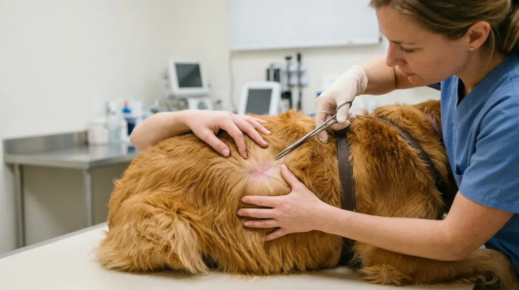
Beyond generalized allergic dermatitis, Golden Retrievers are uniquely predisposed to two breed-specific dermatological conditions that require precise diagnostic differentiation: acute moist dermatitis (hot spots) and congenital ichthyosis.
| Diagnostic Parameter | Acute Moist Dermatitis (Hot Spots) | Golden Retriever Ichthyosis (PNPLA1) | Allergic Atopic Dermatitis |
|—|—|—|—|—|
| Primary Etiology | Rapid self-trauma over wet, matted undercoat with bacterial proliferation | Autosomal recessive genetic defect in lipid metabolism | Polygenic skin barrier defect with immune hypersensitivity |
| Primary Lesion Appearance | Well-demarcated, erythematous, moist, weeping, ulcerated plaque | Large, soft, non-inflammatory brownish-grey to black adherent scales | Diffuse erythema, papules, excoriations, salivary staining |
| Onset Velocity | Rapid development over 4 to 12 hours | Slow, gradual progression appearing in young puppyhood | Gradual onset between 6 months and 3 years |
| Itch Intensity | Extreme localized pain and frenzied scratching | Minimal to mild baseline pruritus without secondary infection | Moderate to severe generalized pruritus across body |
| First-Line Management | Hair clipping, antiseptic drying washes, topical/systemic antibiotics | Emollient rinses, spot-on ceramides, keratolytic shampoos | Systemic targeted biologics (Cytopoint, Apoquel), allergy control |
Acute Moist Dermatitis (Hot Spots)
Acute moist dermatitis, clinically termed pyotraumatic furunculosis, represents a dermatological emergency common in heavy-coated water retrievers. Hot spots begin when localized cutaneous irritation, such as an insect bite, mild ear infection, or wet undercoat matting, prompts frantic licking, scratching, and chewing. Within hours, self-inflicted mechanical trauma shears the superficial epithelium, allowing resident Staphylococcus bacteria to multiply rapidly in the warm, exudative wound bed.
The lesion quickly transforms into a hairless, intensely painful, weeping red plaque that expands outwards across adjacent skin. Hot spots frequently develop along the lateral neck, base of the ears, thighs, and lumbar spine. Because the dense double coat mats over the weeping discharge, it seals in moisture and accelerates tissue maceration. Severe localized pain and acute distress can cause dramatic behavioral changes and muscle trembling, distinct from systemic chills or why French Bulldogs experience shaking during generalized anxiety.
Golden Retriever Ichthyosis
Golden Retriever ichthyosis is a hereditary non-epidermolytic cornification disorder caused by an autosomal recessive mutation in the PNPLA1 (patatin-like phospholipase domain containing 1) gene, as characterized in a veterinary study on canine nonepidermolytic ichthyosis. The PNPLA1 enzyme plays an essential role in synthesizing omega-hydroxy ceramides, which bind the lipid envelope of the epidermal stratum corneum.
When a Golden Retriever inherits two copies of this mutated gene (one from each parent), abnormal keratinization results in large, soft, brownish-grey to black fish-like scales that adhere to the skin of the trunk, abdomen, and flanks. Unlike allergic dermatitis, pure ichthyosis is non-inflammatory and typically causes minimal to no itching. However, because the defective stratum corneum compromises natural antimicrobial defenses, dogs with ichthyosis frequently develop secondary bacterial or Malassezia yeast dermatitis, which introduces acute secondary pruritus.
Identifying Symptoms: How and Where Itchiness Presents
Recognizing localized anatomical symptom patterns enables owners to distinguish between environmental allergies, flea infestations, food sensitivities, and secondary microbial infections. The American Kennel Club’s veterinary guide to dog allergies emphasizes that canine allergic disease manifests across distinct anatomical target zones.
| Anatomical Region | Primary Clinical Symptoms & Lesions | Common Behavioral Signs |
|---|---|---|
| Ears and Periocular Area | Bilateral otitis, erythema of pinnae, waxy brown discharge, yeasty odor | Head shaking, pawing at ears, rubbing face on carpets and furniture |
| Paws and Interdigital Spaces | Erythema between toes, salivary porphyrin staining, secondary yeast infections | Compulsive licking, chewing, and biting at feet and nail beds |
| Ventral Abdomen and Groin | Macular redness, papules, pustules, chronic hyperpigmentation, lichenification | Scratching flanks, rolling on rough surfaces, grooming belly raw |
| Dorsum and Tail Base | Alopecia, broken hairs, punctate crusts, visible flea dirt (digested blood) | Frenzied biting at lower back, sudden agitation, tail chasing |
Ears and Facial Region
Canine ear canals and nasal passages are lined with modified epidermal tissue that reacts aggressively to inhaled aeroallergens. In atopic Golden Retrievers, allergic mucosal swelling and chronic inflammation often provoke frequent head shaking, muzzle rubbing, and loud reverse sneezing. While distinct structural airway features explain why Pug dogs snort during normal breathing, sudden snorting or frantic head shaking in a retriever typically indicates severe allergic nasopharyngeal irritation, secondary otitis externa, or inhaled foreign plant awns.
Paws and Interdigital Webbing
Compulsive paw licking and chewing is a hallmark sign of environmental atopy and adverse food reactions. Allergens absorbed percutaneously through foot pads induce intense interdigital pruritus. As your Golden Retriever licks their feet continuously, porphyrin pigments in canine saliva oxidize upon exposure to air, staining light cream-to-golden fur a distinctive rusty reddish-brown. Over time, chronic friction and moisture cause interdigital furunculosis, painful cysts, and secondary yeast infections between the toes, mirroring compulsive oral behaviors such as why Pug dogs lick so much during periods of skin discomfort.
Ventral Abdomen, Groin, and Axillae
The thinly haired skin along the belly, inner thighs, and armpits readily absorbs environmental pollens when dogs walk through grass. Acute inflammation presents as bright pink macular erythema and small papules. With chronic scratching, repeated self-trauma prompts the skin to defend itself through hyperpigmentation (darkening to black or charcoal grey) and lichenification, where the dermis thickens and develops deeply wrinkled, leathery textures commonly referred to as “elephant skin.”
Lumbosacral Dorsum and Tail Base
The dorsal lumbar spine, base of the tail, and caudal thighs form the classic “flea triangle.” When flea saliva antigens trigger acute hypersensitivity, dogs direct intense chewing and biting toward their lower back. Characteristic findings include thinning hair, broken guard hairs, punctate crusting, and fine black granular “flea dirt” (digested blood feces) that turns reddish-brown when placed on a wet paper towel.
How Veterinarians Diagnose the Root Cause
Because environmental atopy, food allergies, parasite infestations, and secondary infections share overlapping clinical signs, veterinarians follow a systematic elimination workflow to establish an accurate diagnosis.
| Diagnostic Step | Clinical Procedure | Primary Target Condition | Diagnostic Timeline & Accuracy |
|---|---|---|---|
| Step 1: In-Clinic Cytology | Skin tape impressions, glass slide smears, and ear swab stains | Bacterial pyoderma (Staphylococcus) and Malassezia yeast overgrowth | Same-day clinic results; highly sensitive for active microbial infection |
| Step 2: Parasite Scraping & Exclusion | Superficial and deep skin scrapings, broad-spectrum isoxazoline trials | Demodex mites, Sarcoptes scabiei, and flea infestations | Immediate microscopic evaluation; 100% rules out ectoparasitic causes |
| Step 3: Strict Elimination Diet Trial | Exclusive prescription hydrolyzed or novel protein diet | Cutaneous Adverse Food Reactions (food allergies) | Several weeks; validated standard for diagnosing dietary allergies |
| Step 4: Environmental Allergy Testing | Intradermal Skin Testing (IDT) or Serum Allergen-Specific IgE Serology | Environmental Atopic Dermatitis (pollen, dust mites, mold) | Formulates targeted allergen-specific immunotherapy (ASIT) |
Step 1: Direct In-Clinic Cytology
The first diagnostic step in any itchy Golden Retriever is assessing surface microflora. Using clear adhesive tape preps, direct skin impression slides, and cotton ear swabs stained with modified Wright-Giemsa stain, the veterinarian examines samples under oil-immersion microscopy. Cytology quantifies the presence of cocci bacteria (Staphylococcus pseudintermedius), rod-shaped bacteria, and budding yeast organisms (Malassezia pachydermatis). Identifying and treating active secondary infections is essential, as microbial overgrowth induces intense pruritus that masks underlying primary allergy patterns.
Step 2: Parasite Exclusion
Veterinarians conduct superficial and deep skin scrapings to check for microscopic mites, including non-contagious Demodex canis residing within hair follicles and highly contagious Sarcoptes scabiei burrowing in the superficial epidermis. Even if live fleas or flea dirt are not visible during physical examination, all allergic workups require initiating strict monthly broad-spectrum isoxazoline parasiticide therapy (such as fluralaner, sarolaner, or afoxolaner) to definitively eliminate flea allergy dermatitis from the diagnostic equation.
Step 3: Strict Elimination Diet Trial
The only scientifically validated method for diagnosing a canine food allergy is a strict elimination diet trial conducted over an extended period of strict dietary control. During this trial, the dog must consume exclusively a veterinary prescription hydrolyzed protein diet (where dietary proteins are enzymatically cleaved into tiny peptides below 3,000 Daltons to evade immune detection) or a veterinary novel protein diet (such as venison, rabbit, or kangaroo) with which the dog has had zero prior exposure.
Over-the-counter commercial “grain-free” or “limited ingredient” pet store foods are clinically inadequate for diagnostic trials due to widespread shared-line manufacturing contamination with undeclared poultry and beef proteins. Furthermore, commercial direct-to-consumer blood, hair, and saliva allergy test kits lack peer-reviewed scientific validity and exhibit high false-positive rates. During the elimination trial, the dog cannot receive table scraps, rawhides, flavored chewable medications, or treats. If gastrointestinal symptoms accompany pruritus, veterinarians assess digestive tolerance, distinguishing food hypersensitivity from acute GI distress like why French Bulldogs keep throwing up after dietary indiscretions. Similarly, identifying carbohydrate or protein malabsorption helps differentiate subtle allergic colitis from benign flatulence such as why French Bulldogs pass excessive gas due to aerophagia.
Step 4: Environmental Atopy Confirmation
If pruritus persists after resolving secondary infections, maintaining strict flea control, and completing an uneventful elimination trial, a clinical diagnosis of Canine Atopic Dermatitis is confirmed. To identify specific environmental culprits, a board-certified veterinary dermatologist performs Intradermal Skin Testing (IDT), injecting tiny amounts of standardized allergen extracts into the dermis and measuring wheal-and-flare reactions, or orders Serum Allergen-Specific IgE Serology. These tests are used exclusively to formulate customized desensitization immunotherapy, not to diagnose food allergies.
Veterinary Treatments and Daily Management for Itchy Goldens
Successfully managing chronic allergic pruritus in Golden Retrievers requires a multimodal therapeutic strategy tailored to the dog’s age, symptom severity, and lifestyle. Rather than relying on broad-spectrum immunosuppressants like oral corticosteroids, modern veterinary medicine uses targeted molecular therapies that control itch pathways with minimal systemic side effects.
| Therapeutic Class | Active Agent / Medication | Primary Biological Mechanism | Speed of Clinical Onset | Administration Route & Frequency | Primary Clinical Indication |
|---|---|---|---|---|---|
| Monoclonal Antibody Biologic | Lokivetmab (Cytopoint) | Binds and neutralizes Interleukin-31 (IL-31) cytokine | 24 to 48 hours | Subcutaneous injection periodically as evaluated by vet | Atopic and allergic pruritus across all canine life stages |
| Targeted JAK-1 Inhibitor | Oclacitinib (Apoquel) | Inhibits Janus kinase-1 (JAK-1) inflammatory enzyme | Rapid | Oral tablet daily as prescribed | Rapid control of acute and chronic allergic itch and redness |
| Topical Antiseptics | Chlorhexidine 2-4% + Ketoconazole 1-2% | Disrupts bacterial and fungal cell membranes | Immediate surface kill; cumulative over weeks | Medicated shampoo bath as directed by vet | Active secondary Malassezia and Staphylococcus infections |
| Barrier Restorative Mousses | Phytosphingosine, Ceramides, Essential Lipids | Restores intercellular lipid matrix of stratum corneum | Ongoing application | Leave-on topical mousse applied regularly | Transepidermal water loss and chronic barrier impairment |
| Allergen Immunotherapy | Custom ASIT formulation (SCIT / SLIT) | Desensitizes immune system via controlled micro-doses | Gradual long-term benefit | Subcutaneous injections or sublingual oral drops as scheduled | Lifelong disease modification for environmental atopy |
| Nutritional Anti-Inflammatory | Marine EPA + DHA Omega-3 Fatty Acids | Competes with arachidonic acid in eicosanoid pathways | Ongoing daily feeding | Daily oral liquid or concentrated soft gel capsule | Adjunctive cutaneous barrier and anti-inflammatory support |
Targeted Systemic Medications
Modern veterinary medicine relies primarily on two targeted therapies:
- Cytopoint (Lokivetmab): An injectable caninized monoclonal antibody designed specifically to mimic natural immune proteins. Cytopoint circulates in the bloodstream and binds directly to Interleukin-31 (IL-31), the neuroimmune cytokine responsible for sending itch signals from inflamed skin to the central nervous system. Because Cytopoint is metabolized through natural protein degradation pathways rather than hepatic or renal filtration, it is safe for puppies of any age and dogs with concurrent liver or kidney conditions. A single injection provides sustained itch relief, with duration depending on the dog’s individual clinical response.
- Apoquel (Oclacitinib): A daily oral medication that inhibits the Janus kinase-1 (JAK-1) enzyme pathway. By blocking JAK-1, Apoquel rapidly shuts down the intracellular signaling of multiple itch-inducing and pro-inflammatory cytokines, including IL-31, IL-2, IL-4, and IL-13. Apoquel provides rapid relief, making it ideal for managing acute seasonal flare-ups and fast itch suppression.
Topical Therapies and Barrier Restoration
Medicated bathing is essential for removing surface allergens, dander, and microbial toxins. Shampoos combining 2% to 4% chlorhexidine gluconate (antibacterial) and 1% to 2% ketoconazole or miconazole (antifungal) clear secondary infections without drying out the coat. For maximum effectiveness, the shampoo lather must remain in contact with the wet skin for a full 10 minutes before being thoroughly rinsed away with cool water.
Between baths, leave-on topical mousses, sprays, and spot-on treatments containing synthetic ceramides, free fatty acids, and phytosphingosine help rebuild the defective epidermal lipid mortar, reducing transepidermal water loss and preventing allergen penetration.
Allergen-Specific Immunotherapy (ASIT)
Allergen-Specific Immunotherapy represents the only therapeutic modality capable of permanently modifying the underlying disease process rather than merely suppressing clinical symptoms. Formulated from the results of intradermal or serological allergy testing, custom immunotherapy delivers gradually increasing micro-doses of your dog’s specific environmental allergens via subcutaneous injections (SCIT) or sublingual oral drops (SLIT). Over time, this controlled exposure gradually trains the immune system to tolerate environmental allergens, significantly reducing or eliminating the need for lifelong daily pharmaceutical medications.
Nutritional Support and Fatty Acids
Supplementing your Golden Retriever’s diet with high-potency marine-derived Omega-3 fatty acids (Eicosapentaenoic Acid [EPA] and Docosahexaenoic Acid [DHA]) provides natural anti-inflammatory benefits. Aim for a combined therapeutic dose of 50 to 75 mg of EPA and DHA per kilogram of body weight daily. These marine lipids incorporate into cell membranes, competing with arachidonic acid and decreasing the production of inflammatory prostaglandins and leukotrienes.
When chronic itching disrupts your dog’s sleep and emotional well-being, pet owners often notice changes in attachment, contrasting baseline canine companionship like why French Bulldogs become clingy with compulsive nighttime restlessness where an allergic dog repeatedly circles or exhibits behaviors like why Pugs dig in blankets to alleviate belly irritation against soft fabric. Managing canine allergies in Golden Retrievers is a lifelong multimodal control strategy rather than a one-time cure. Combining targeted biologic therapy (Cytopoint) or JAK inhibitors (Apoquel) with strict coat-drying protocols and skin barrier supplementation yields significantly better long-term comfort with fewer secondary infections than relying on antihistamines or periodic steroid bursts.
Original Value & Breed-Specific Analysis: The “Swimmer’s Coat Trap”
Golden Retrievers possess an innate affinity for water, but their water-repellent double coat presents a unique dermatological paradox known among veterinary dermatologists as the “Swimmer’s Coat Trap.”

| Stage | Physical Event | Cutaneous Impact | Microclimate & Infection Risk |
|---|---|---|---|
| 1. Water Infiltration | Dense undercoat becomes fully saturated during swimming | Traps moisture against dermis beneath oily guard hairs | Outer coat collapses, preventing natural air evaporation |
| 2. Contaminant Deposition | Microscopic silt, algae, pond bacteria, and chlorine settle in fur | Chemical and biological agents contact epidermal barrier | Trapped particulates dissolve natural protective lipid film |
| 3. Microclimate Formation | Trapped moisture combines with 101-102.5 °F canine body heat | Keratinocytes soften through continuous tissue maceration | Creates an ideal warm, dark, anaerobic incubation chamber |
| 4. Bacterial Proliferation | Commensal Staphylococcus bacteria multiply explosively | Triggers acute dermal pain, severe itching, and self-trauma | Rapidly expands into full weeping hot spot within 12 to 24 hours |
While coarse guard hairs shed surface water, the ultra-dense undercoat absorbs moisture like a sponge when fully submerged. When a Golden Retriever exits a lake, swimming pool, river, or ocean, the outer guard hairs collapse over the wet underfur, sealing in moisture against warm body tissue.
Furthermore, outdoor water sources contain organic silt, blue-green algae, fungal spores, and microscopic bacteria, while treated swimming pools contain harsh chlorine and algaecides. As the trapped water sits against the skin for hours, these chemical and biological contaminants dissolve the skin’s protective lipid film. The stratum corneum becomes macerated (softened and weakened by continuous wetness), lowering local tissue immunity and allowing resident Staphylococcus pseudintermedius bacteria to trigger rapid pyotraumatic furunculosis (hot spots) within 12 to 24 hours.
The 4-Step Post-Swimming Prevention Protocol
To protect your water-loving Golden Retriever from moisture-induced skin breakdown, implement this standardized post-swim care protocol:
- Immediate Complete Freshwater Rinse: Immediately after swimming in lakes, rivers, ocean salt water, or chlorinated pools, thoroughly hose down your Golden Retriever with clean, fresh tap water. Ensure the water stream penetrates beneath the topcoat to flush out sand, sediment, chlorine, and organic bacteria trapped against the skin.
- Forced-Air High-Velocity Drying: Towel drying alone is insufficient for a water-logged Golden Retriever double coat. Use a dedicated high-velocity forced-air pet dryer to separate the coat and blow moisture outward from the skin level. Always use cool or warm airflow settings; never use high heat, which damages delicate epidermal proteins and inflames sensitive skin.
- Prophylactic Astringent Ear Cleaning: Following every swimming session, instill a veterinary-approved drying ear cleaner containing salicylic acid, acetic acid, or boric acid into both ear canals. Gently massage the base of the ear for 30 seconds to allow the astringent solution to dissolve trapped water and wax, then let your dog shake their head before wiping excess fluid with a clean cotton pad.
- Strategic Cutaneous Spot-Checks: Once the coat is completely dry, physically part the fur with your fingers along high-risk anatomical zones: beneath the collar on the neck, behind the ear leather, along the groin folds, and at the base of the tail. Catching a localized patch of moist redness early allows for prompt topical astringent disinfection before it expands into a painful hot spot. Adhering to these steps ensures comprehensive essential dog care protocols that safeguard your retriever’s active lifestyle.
Frequently Asked Questions About Itchy Golden Retrievers
What are golden retrievers most allergic to?
Golden Retrievers are most frequently allergic to environmental aeroallergens (such as grass, weed, and tree pollens, house dust mites, and fungal mold spores), followed by flea saliva antigens and common dietary animal proteins (particularly chicken, beef, dairy, and egg). Environmental atopic dermatitis is the most prevalent allergic disease diagnosed in the breed.
What home remedy can I give my golden retriever for itching?
Safe home management includes bathing your dog with a gentle, hypoallergenic colloidal oatmeal shampoo, applying cool water compresses to localized hot areas, and wiping paws with a damp cloth after outdoor walks to physically remove surface pollen. However, home remedies only provide temporary surface soothing and will not resolve underlying bacterial pyoderma, yeast dermatitis, flea allergies, or systemic atopy, which require veterinary medical intervention.
Is Cytopoint or Apoquel better for a golden retriever?
Neither medication is universally superior; the ideal choice depends on your dog’s age, symptom pattern, and lifestyle. Cytopoint is an injectable monoclonal antibody administered periodically under veterinary guidance that specifically neutralizes the itch-inducing IL-31 cytokine with minimal hepatic or renal involvement, making it suitable for dogs of any age or those with sensitive stomachs. Apoquel is a daily oral tablet that inhibits the JAK-1 enzyme, rapidly suppressing both itching and active inflammatory redness.
Can diet alone cure my golden retriever’s itchy skin?
Dietary modification will only resolve itching if your Golden Retriever suffers from a true cutaneous adverse food reaction, which accounts for only 10% to 15% of all allergic canine skin cases. If your dog has environmental atopic dermatitis, flea allergy dermatitis, or congenital ichthyosis, changing dog food brands will not stop the scratching, and targeted medical management will remain necessary.
How do I know if my golden retriever has hot spots or regular allergies?
Allergies generally present as diffuse, widespread itching characterized by pink skin, paw chewing, facial rubbing, and recurrent ear inflammation that fluctuates over time. In contrast, a hot spot (acute moist dermatitis) is an acute, localized, intensely painful, wet, and rapidly expanding skin sore that can develop from a tiny scratch into an ulcerated, weeping lesion several inches wide in just a few hours.



Leave a Reply