
Table of Contents
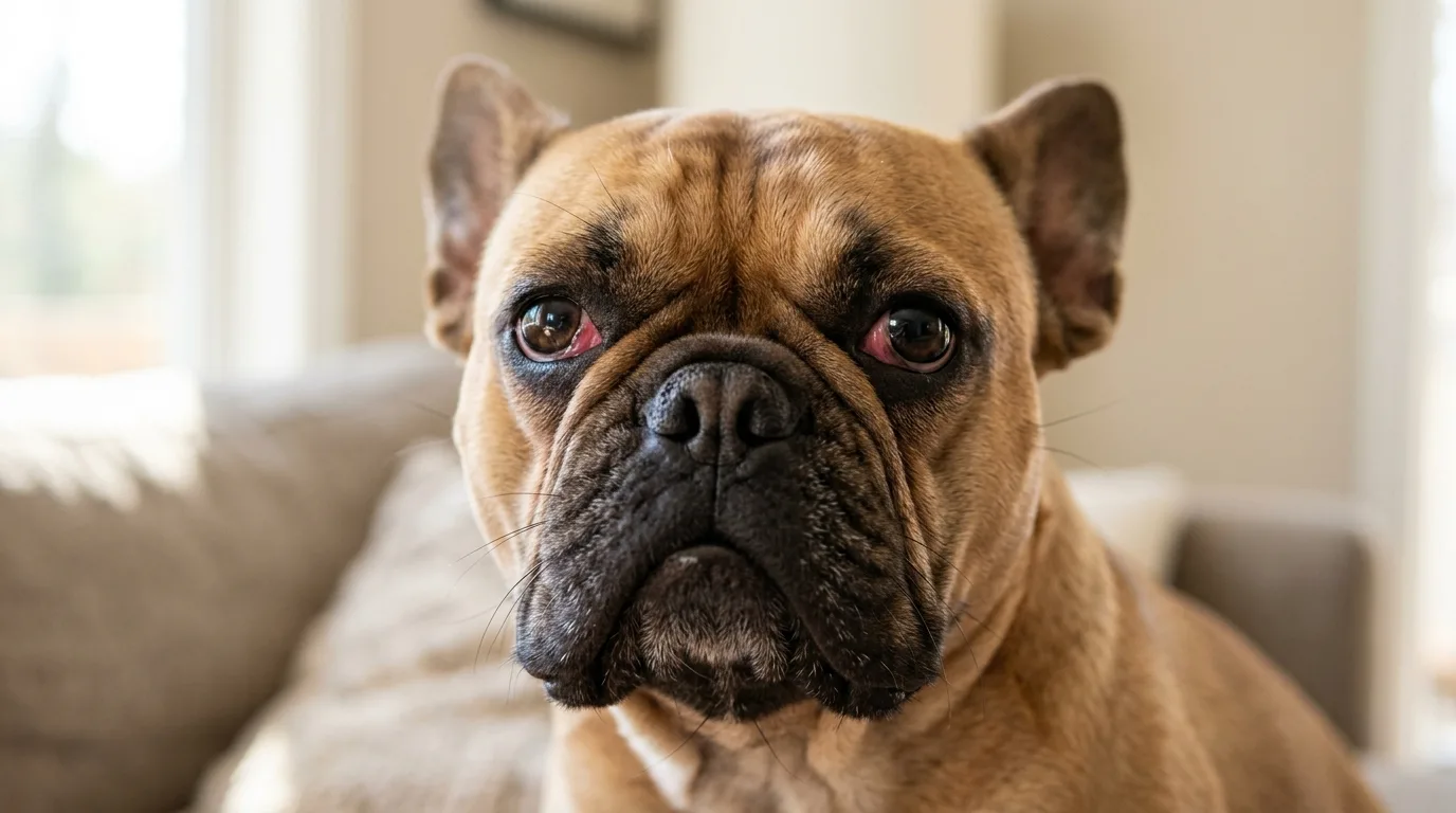
If you find yourself wondering, "why are my French bulldogs eyes red," the cause is often a mild irritant, but it can also signal painful disorders like a corneal ulcer, tear deficiency, or glaucoma. Because Frenchies have prominent facial features, any redness paired with squinting, cloudiness, a held-shut lid, or bulging requires urgent veterinary assessment.
- Redness is a symptom of many conditions, not a diagnosis.
- French Bulldogs are at high risk for corneal ulcers due to their facial conformation.
- Cherry gland prolapse appears as a pink lump at the inner corner and needs surgical repositioning.
- Never use human drops or leftover prescriptions without a vet's approval.
- Pain, sudden cloudiness, or bulging are immediate veterinary emergencies.
Why French Bulldogs are prone to ocular irritation
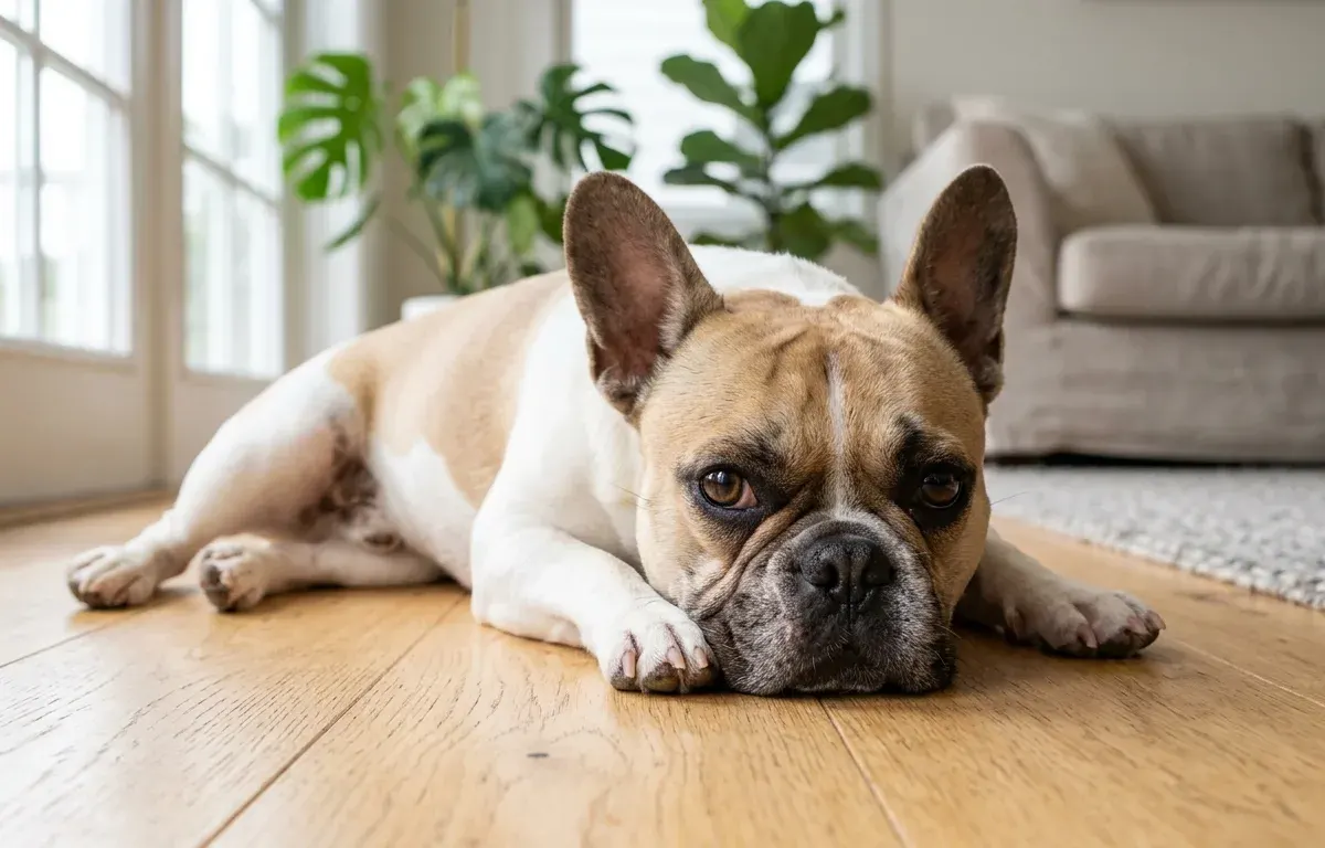
French Bulldogs are brachycephalic dogs with flat faces and prominent features. This anatomical structure directly affects their optical health. Their shortened muzzles, larger lid openings, exposed sclera, and nasal folds increase the risk of surface injuries. For context on what makes a French Bulldog a brachycephalic breed, their skull shape limits the protective housing of the socket.
These traits expose the cornea to environmental damage. Researchers also note that brachycephalic dogs have reduced corneal nerve density, meaning they might not feel a minor scratch until it worsens. The Royal Veterinary College (RVC) Ophthalmology Service notes that brachycephalic breeds represent about 25 percent of their canine patients, many presenting with deep or melting corneal ulcers. Understanding the breed’s original purpose and history helps explain how structural changes over time led to these modern health realities.
Are French Bulldogs prone to inherited ocular disorders?
Veterinary specialists recognize several inherited conditions in the breed. These include entropion (inward-rolling lids), distichiasis (abnormal lashes), prolapsed third-lid gland, and poor tear production. The presence of these known risks lowers the threshold for a thorough examination. Discoloration does not automatically mean the dog has a genetic defect, but the breed’s predisposition makes a proper veterinary check necessary.
Common causes of ocular redness in French Bulldogs
Inflammation can involve the conjunctiva, the corneal surface, the lids, or deeper internal structures. The symptoms overlap significantly.
| Possible category | What an owner may notice | Why home diagnosis is unreliable | Urgency |
|---|---|---|---|
| Environmental irritants | Watering, mild rubbing | Overlaps with early ulcers | Contact vet if it persists |
| Conjunctivitis | Redness, swelling, discharge | Could be secondary to a bigger issue | Needs diagnosis |
| Keratoconjunctivitis sicca (KCS) | Thick mucus, recurring redness | Cannot be measured at home | Vet visit required |
| Gland prolapse | Pink or red lump in corner | Obvious, but needs surgical care | Schedule a visit |
| Corneal ulcers | Squinting, pain, cloudiness | Fast progression to rupture | Urgent to emergency |
| Glaucoma/Uveitis | Severe pain, pupil changes | Vision loss happens quickly | Immediate emergency |
Environmental irritation, allergy, or a foreign object
Dust, smoke, pollen, and foreign material frequently irritate the surface and cause redness, tearing, blinking, or rubbing. Allergen-driven follicular conjunctivitis is also common in young dogs. However, these signs look identical to the early stages of a corneal injury or reduced tear production. Persistent symptoms require professional evaluation rather than assuming the dog just has allergies.
Conjunctivitis is a finding, not always the root cause
Conjunctivitis is the inflammation of the tissue lining the lids and covering the white part of the sclera. According to VCA Hospitals on conjunctivitis, typical signs include redness, swelling, discharge, and squinting. It arises from allergies, infections, tear deficiencies, lash abnormalities, ulcers, uveitis, glaucoma, or trauma. Treating this inflammation requires finding and addressing the underlying trigger.
Tear deficiencies and lid or lash disorders behind recurring redness
Keratoconjunctivitis sicca (KCS) occurs when tear production drops. The area becomes red, painful, and irritated, producing a thick yellowish mucoid discharge. VCA Hospitals on dry eye confirms this lack of lubrication can drive recurrent conjunctivitis or corneal ulcers. Inward-rolling lids or abnormal lashes can also rub the cornea continuously, causing secondary inflammation.
Gland prolapse: a red lump at the inner corner
A prolapsed gland of the third eyelid usually appears as a round pink swelling at the inner lower corner. Because this gland produces 30 to 50 percent of the tears, veterinary sources like VCA Hospitals on cherry eye recommend surgically repositioning it. Removing the gland increases the risk of the dog developing severe dryness later in life.
Corneal ulcers, uveitis, and glaucoma need urgent exclusion
Discoloration can indicate a serious emergency. Corneal ulcers are painful, causing squinting, tearing, and rubbing, and they can rapidly deepen toward a rupture. Sudden closed-angle glaucoma brings severe pain, redness, a cloudy cornea, and vision loss. Uveitis also reddens the sclera.
Note: A painful or abnormal-looking condition requires prompt veterinary care, not trial-and-error drops. Glaucoma and severe ulcers threaten vision and require immediate medical intervention.
One side or both? Read the pattern carefully
A single affected side often fits a localized problem like trauma, a foreign body, a corneal injury, or a unilateral prolapsed gland. Both sides turning red can point to allergies, tear deficiency, infection, or bilateral structural issues. Neither pattern is diagnostic on its own. Any pain or vision change overrides pattern-watching. For instance, unusual shaking or trembling in a French Bulldog alongside facial pain suggests significant distress and demands urgent care.
| Pattern | Possible causes | What it does NOT prove |
|---|---|---|
| One side affected | Trauma, foreign body, ulcer, unilateral prolapsed gland | Does not confirm a minor scratch |
| Both sides affected | Allergies, KCS, systemic infection, bilateral structural issues | Does not confirm a harmless allergy |
When redness needs a vet urgently
Seek same-day advice for sudden discoloration accompanied by squinting, repeated blinking, pawing, or discharge. Go to an emergency vet immediately for a held-shut or intensely painful lid, a cloudy cornea, visible bulging, a changed pupil, suspected trauma, or sudden vision loss.
The MSD Veterinary Manual warns that proptosis (the globe coming out of its socket) is an ocular emergency where the surface dries out and vision fails without immediate surgical replacement. Owners should remember that when a Frenchie’s repeated vomiting needs a vet, sudden severe symptoms always warrant prompt contact. While lethargy can happen, assessing whether French Bulldogs sleep a lot normally should not delay a vet visit if the dog is hiding due to discomfort.
| Urgency Level | Symptoms to watch for | Action required |
|---|---|---|
| Emergency now | Held-shut lid, severe pain, cloudy cornea, bulging, vision loss | Go to the nearest emergency vet immediately |
| Contact vet today | Squinting, repeated blinking, new discharge, pawing | Call your primary vet for a same-day appointment |
| Monitor | Brief redness after a known minor irritant with no pain | Watch closely, call if it persists |
What NOT to put on the dog’s face
Do not use human redness-relief drops, leftover prescriptions, or steroid medication unless a vet performs an examination and prescribes it for the current problem. Medication that worked for a past episode can cause harm if the underlying cause is different. Steroid solutions, in particular, can rapidly worsen an undiagnosed corneal ulcer.
Safe steps before the veterinary visit
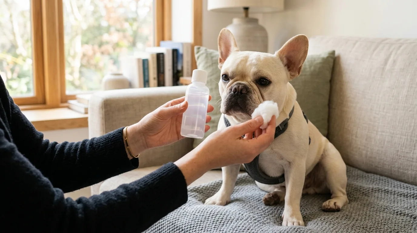
Stop the dog from rubbing the area. An Elizabethan collar (cone) prevents self-trauma. Avoid all unprescribed drops and phone a vet for direction. If the vet advises it, or if a minor surface irritant is plainly visible, flush the affected area only with sterile saline made for irrigation.
Limit the dog’s physical activity. Consider safe activity limits and exercise for a French Bulldog to prevent further trauma during rough play. Avoid swimming, as why French Bulldogs need water safety extends to keeping contaminated water out of an irritated socket.
How a veterinarian finds the cause
A vet examines the cornea, lids, lashes, tear ducts, and surrounding structures. They often use a fluorescein stain to reveal corneal injuries or ulcers, a Schirmer test to measure tear production, and tonometry to measure intraocular pressure for glaucoma screening.
| Diagnostic Test | What it checks | Why it matters |
|---|---|---|
| Fluorescein stain | The corneal surface | Identifies scratches and ulcers |
| Schirmer test | Tear production levels | Diagnoses KCS |
| Tonometry | Intraocular pressure | Screens for glaucoma and uveitis |
Routine care at home has limits. Knowing how to safely clean French Bulldog ears is standard grooming, but assessing ocular pain requires professional medical tools.
Reducing repeat irritation after the diagnosis
Prevention depends on the specific diagnosis. After the vet identifies the cause, owners can keep the facial folds clean as directed, prevent rubbing, and limit exposure to windblown debris. Attend all recommended rechecks, especially if the dog has tear deficiencies, lid abnormalities, or recurring corneal problems. Managing a Frenchie’s health requires proactive monitoring. Just like understanding IVDD in French Bulldogs, recognizing their baseline optical health helps owners spot deviations early before they escalate into emergencies.
FAQ about French Bulldog ocular redness
Will mild redness clear up on its own?
Brief discoloration after a known minor irritant may settle, but you cannot confirm the cause safely at home. Get veterinary advice if the redness persists, returns, or comes with discharge, rubbing, squinting, cloudiness, or a behavior change.
Is a prolapsed gland the same as being bloodshot?
No. A prolapse involves the third lid’s gland, appearing as a pink or red mass at the inner corner. Being bloodshot simply involves inflamed blood vessels on the scleral surface.
Why is the dog holding one side shut?
Keeping the lid clamped or intense squinting signals significant pain. This occurs with corneal injuries, foreign bodies, severe inflammation, or pressure-related diseases like glaucoma. Treat it as an urgent reason to contact a veterinarian.
Can these symptoms be diagnosed from a photo?
No. A photo helps document progression for the veterinary team, but a vet cannot measure tear production, check intraocular pressure, stain the cornea, or look at internal structures using only a picture.


Leave a Reply