

Table of Contents
Epidemiological data on how common Pug Dog Encephalitis is indicates that PDE affects an estimated 1.0% to 1.2% of all Pugs over their lifetime. While rare across the overall breed population, it is the predominant intracranial brain disorder in Pugs, accounting for approximately 81% of fatal brain disease cases evaluated in neurology referral centers. Medically designated as necrotizing meningoencephalitis (NME), the condition is a severe, non-contagious autoimmune disorder driven by genetic risk markers located on canine chromosome 12.
- Lifetime prevalence is estimated at 1.0% to 1.2% across the overall Pug population.
- NME represents approximately 81% of fatal intracranial neurological disorders in Pugs.
- The disease is hereditary and autoimmune, associated with Dog Leukocyte Antigen class II markers.
- PDE is strictly non-contagious and cannot spread through contact, saliva, or air.
- Preventive DNA testing through blood or cheek swabs categorizes breeding risk before mating.
- Clinical episodes cause acute pain from intracranial pressure, requiring urgent veterinary neurology care.
What is pug dog encephalitis?
Pug Dog Encephalitis (PDE) is an aggressive, idiopathic inflammatory disease targeting the central nervous system of dogs. In clinical veterinary medicine, PDE is classified as necrotizing meningoencephalitis (NME). This condition is characterized by extensive necrosis and non-suppurative inflammation focused predominantly in the cerebral cortex and leptomeninges of young and middle-aged Pugs. Unlike infectious neurological conditions caused by viral, bacterial, fungal, or protozoal pathogens, PDE is a sterile autoimmune disease in which the dog’s immune system mistakenly attacks its own brain tissue.
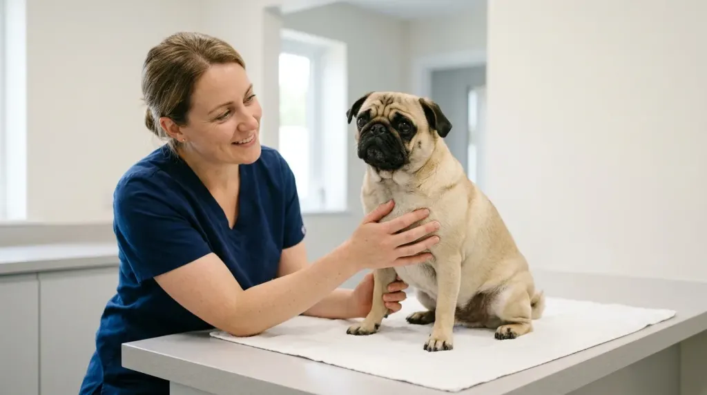
Although necrotizing encephalitis occurs in other small toy breeds such as Maltese and Chihuahuas, the disease displays unique genetic and pathological features in Pugs. Familiarity with what a Pug dog looks like and understanding normal breed anatomy helps owners spot early physical abnormalities, including subtle changes in facial symmetry, head carriage, and gait coordination. Historically, veterinary literature referred to this disorder exclusively as PDE because of its disproportionately high incidence in the breed. Today, veterinary neurologists recognize NME as the broader pathological classification while continuing to study the Pug as the classic model of genetic autoimmune encephalitis.
How common is pug dog encephalitis?
Across the general pet Pug population, Pug Dog Encephalitis is relatively uncommon, with large-scale breed health surveys and kennel registries estimating a lifetime mortality prevalence between 1.0% and 1.2%. This figure indicates that more than 98% of Pugs will never develop the condition during their lifetime. However, within specialized veterinary neurology settings, the disease represents the overwhelming majority of serious brain disorders seen in the breed.
In a retrospective multi-center study published by veterinary researchers, NME accounted for 81% of all Pugs presented with necropsy-confirmed intracranial lesions. This contrast between low general population frequency and high representation in neurology clinics highlights why PDE is both an uncommon life event for average owners and a critical diagnostic priority for veterinarians evaluating neurological symptoms in Pugs.
| Population Cohort | Estimated Prevalence / Proportion | Data Source and Study Context |
|---|---|---|
| General Pug Population | 1.0% to 1.2% lifetime prevalence | Breed health surveys and veterinary epidemiological registries |
| Pugs with Confirmed Fatal Intracranial Disease | 81% of evaluated cases | Multi-center veterinary neurology retrospective cohort |
| High-Risk Allele Frequency (DLA-DPB1) | ~25.7% risk allele frequency | Breed-wide canine genomic diversity surveys |
| Homozygous High-Risk Genotype (S/S) | ~7.4% of tested Pugs | Susceptible homozygous status in commercial DNA screening |
Is pug dog encephalitis contagious?
Pug Dog Encephalitis is completely non-contagious. Because the underlying mechanism is an internal immune system malfunction governed by inherited genetic susceptibility, the disease cannot spread from one dog to another. Transmission through physical contact, shared food and water bowls, saliva, respiratory droplets, or environmental surfaces is impossible.
Owners managing multiple dogs do not need to institute quarantine protocols when a household Pug receives an NME diagnosis. Healthy companion pets and human family members face zero infection risk. Disease prevention strategies focus entirely on selective breeding practices informed by DNA marker status rather than biosecurity or isolation measures.
Is pug dog encephalitis hereditary?
PDE has a proven hereditary foundation linked to the Dog Leukocyte Antigen (DLA) class II complex located on canine chromosome 12. Specifically, researchers identified the CFA12:2605517delC marker in the DLA-DPB1 gene as a major genetic risk factor. Pugs that inherit two copies of this risk marker (homozygous high-risk, designated S/S) have a 12.75-fold increased likelihood of developing clinical NME compared to dogs with zero risk copies (homozygous low-risk, N/N).
Despite this clear genetic association, PDE follows a complex, non-Mendelian inheritance pattern. Approximately 7.4% of Pugs carry the homozygous S/S genotype, yet only about 1.0% to 1.2% develop active encephalitis. This discrepancy demonstrates that inheriting the genetic risk variant is necessary but not solely sufficient to trigger disease onset. Additional secondary modifier genes, epigenetic influences, and environmental triggers are required for the autoimmune reaction to manifest. Prospective owners investigating where to buy a Pug dog should request official parental DNA health testing certificates from responsible breeders before acquiring a puppy.
What causes pug dog encephalitis?
The exact initiating trigger for PDE remains the subject of ongoing scientific inquiry, but the core pathological process is established as a sterile autoimmune cascade. Extensive polymerase chain reaction (PCR) investigations of cerebrospinal fluid and affected brain tissues consistently fail to detect common canine neurotropic viruses (including canine distemper virus, herpesvirus, adenovirus, parvovirus, and rabies), confirming that NME is an immune-mediated disorder rather than an infectious disease.
During active disease, autoreactive T-lymphocytes cross the blood-brain barrier and mount an aggressive attack against central nervous system antigens. Cerebrospinal fluid analysis reveals autoantibodies specifically targeting glial fibrillary acidic protein (GFAP), a structural protein produced by astrocytes in cerebral white and grey matter. This targeted autoimmune assault causes severe parenchymal necrosis, cerebral edema, loss of cortical tissue, and secondary elevation of intracranial pressure.
How is pug dog encephalitis diagnosed?
Evaluating a Pug for PDE involves two distinct clinical situations: proactive genetic screening in healthy dogs and diagnostic evaluation in symptomatic patients.
-
Preventive Genetic DNA Screening: Performed using a cheek swab or whole blood sample submitted to the UC Davis Veterinary Genetics Laboratory. The assay evaluates the DLA class II risk markers and returns one of three genotypes:
- Low Risk (N/N): Carries two normal copies. Minimal probability of developing NME; safe to breed to any genotype.
- Carrier / Moderate Risk (N/S): Carries one normal and one risk copy. Low individual probability of disease, but transmits the risk marker to 50% of offspring.
- High Risk (S/S): Carries two risk copies. Possesses a 12.75-fold elevated lifetime risk of developing clinical NME and transmits the risk marker to all offspring. High-risk dogs should never be mated with another S/S or N/S dog.
-
Clinical Diagnostic Investigation: In dogs exhibiting active neurological signs, presumptive antemortem diagnosis and exclusion of other etiologies rely on multi-modal veterinary testing:
- Magnetic Resonance Imaging (MRI): Demonstrates asymmetric, multifocal T2-weighted and FLAIR hyperintensities in the cerebral cortex and subcortical white matter, often accompanied by loss of grey-white matter differentiation and mass effect.
- Cerebrospinal Fluid (CSF) Analysis: Demonstrates marked mononuclear or mixed-cell pleocytosis (elevated nucleated white blood cell count) alongside elevated microprotein concentrations.
- Systemic Blood and Metabolic Testing: Complete blood count, serum biochemistry, and infectious disease PCR panels exclude systemic infections, hepatic encephalopathy, metabolic disturbances, and nutritional deficiencies.
- Histopathological Confirmation (Brain Biopsy or Post-Mortem Necropsy): Histopathological examination of affected brain tissue revealing necrotizing cortical lesions and lymphoplasmacytic perivascular cuffing remains the definitive gold standard for confirmation, obtained via specialized antemortem brain biopsy or post-mortem necropsy.
Is pug dog encephalitis painful?
Pug Dog Encephalitis causes substantial physical discomfort and distress during acute and progressive phases. The severe inflammation and subsequent necrosis of brain tissue lead to cerebral edema and elevated intracranial pressure. Affected dogs frequently display head pressing, a recognized neurological sign where the dog compulsively pushes its head against walls, furniture, or floors to alleviate severe intracranial headache.
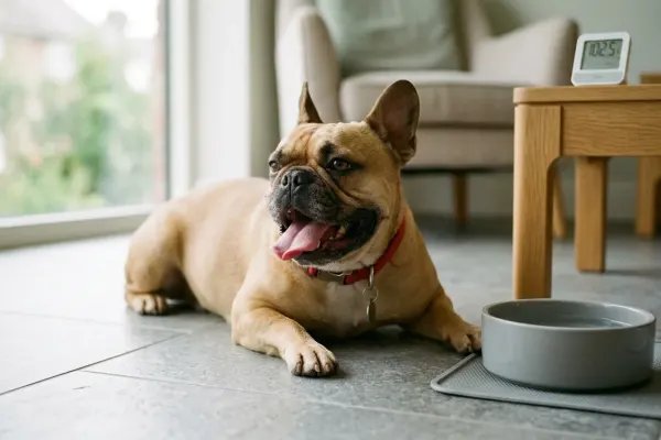
In addition, dogs with NME frequently suffer from cervical hyperesthesia, characterized by intense neck stiffness, muscular spasms, and vocalization upon gentle handling. Differentiating between normal resting behaviors and pathological distress is critical, as severe central nervous system inflammation causes persistent discomfort, compulsive restlessness, and agitation rather than typical relaxed lounging.
Common clinical signs reflecting neurological impairment and acute distress include:
- Generalized tonic-clonic or focal seizures that escalate in frequency and severity
- Compulsive pacing, circling in one direction, and severe disorientation
- Head pressing against solid surfaces and marked neck pain upon flexion
- Sudden vision loss, dilated non-responsive pupils, and altered awareness
- Severe ataxia, weakness in all four limbs (tetraparesis), or acute collapse
Geographic and epidemiologic context
Canine inflammatory central nervous system disorders vary by breed, age, and geographical region. In North America, Europe, and Japan, necrotizing meningoencephalitis is recognized as a breed-predisposed disorder concentrated in Pugs, Chihuahuas, and Maltese. In the Journal of Veterinary Internal Medicine retrospective study, Dr. Jonathan M. Levine and fellow investigators demonstrated that the median age at onset for Pugs with NME is 18 months, with females showing a higher frequency of diagnosis than males.
Epidemiological evaluations indicate that clinical onset occurs most commonly between 6 months and 7 years of age. In younger dogs presenting within this typical window, early subtle signs like nocturnal agitation or compulsive pacing can be easily overlooked; understanding what causes a Pug dog to keep moving and not lay down helps owners spot neurological restlessness before advanced seizures develop. Conversely, while cases in geriatric dogs over 9 years of age have been documented, late-onset neurological signs require careful differentiation from vascular events, intracranial neoplasia, or degenerative cognitive changes seen when Pugs are considered senior dogs. Retrospective clinical data confirms that while Pug Dog Encephalitis has a modest overall lifetime incidence of 1.0% to 1.2%, its concentration among young Pugs presented with sudden-onset forebrain signs makes it the primary differential diagnosis in emergency neurology settings. Genetic screening provides practical breeding guidance, but veterinarians emphasize that clinical symptoms require immediate advanced neuro-imaging and immunosuppressive therapy rather than reliance on DNA status alone.
Note: A high-risk S/S genetic test result is not a clinical diagnosis and does not mean a dog is currently sick. Only an estimated one in seven S/S dogs develops active clinical disease. Never make medical treatment decisions solely on the basis of a preventive DNA test.
Interpreting the data for pug owners
Epidemiological figures put the condition in perspective: because lifetime risk in the general breed population stays around 1.0% to 1.2%, the vast majority of Pugs never experience encephalitis. Routine veterinary wellness checks and structured daily habits for taking care of a Pug dog provide the best baseline for noticing sudden changes in gait or demeanor early. Owners can also distinguish harmless behaviors, like why a Pug dog digs in blankets before curling up to sleep, from pathological pacing, constant circling, or painful neck guarding that warrants emergency attention.

When clinical NME is confirmed, treatment focuses on halting autoimmune tissue destruction with aggressive immunosuppressive protocols and controlling seizures with targeted anticonvulsants. Standard medical regimens combine high-dose oral glucocorticoids such as prednisolone with secondary agents like cytosine arabinoside, cyclosporine, or leflunomide. Although the overall prognosis remains guarded, with retrospective studies documenting a historical mean survival time of 93 days (median 44 days, range 1 to 680 days), prompt multi-agent therapy enables responsive dogs to achieve quality clinical remissions lasting 12 to 24 months or longer.
Summary of current evidence and research gaps
Scientific research over the past two decades has clarified the genetic architecture and immunopathology of Pug Dog Encephalitis. Key established facts include the identification of DLA class II susceptibility markers on chromosome 12, the demonstration of GFAP-directed autoantibodies, and the documentation of characteristic MRI and CSF abnormalities.
Alongside genetic research, holistic canine health studies highlight the role of metabolic balance and nutritional management in supporting overall health. Providing a high-quality, nutrient-dense diet for Pug dogs supports organ function and immune stability across every stage of life. Evaluating overall wellness alongside benchmarks for how long Pug dogs live reinforces the importance of responsible breeding programs aimed at reducing risk allele frequencies while preserving overall genetic diversity in the Pug breed.
Frequently asked questions about pug dog encephalitis
At what age do Pugs usually develop PDE?
Pugs typically develop clinical signs of PDE between 6 months and 7 years of age, with the peak onset occurring around 18 months. Rare cases have been documented in dogs under 4 months or over 9 years old.
Can male Pugs get Pug Dog Encephalitis?
Yes, male Pugs can develop PDE. While some epidemiological surveys show a slightly higher prevalence in females, male Pugs carrying high-risk DLA class II genetic markers remain fully susceptible to the disease.
Can PDE be cured?
No, PDE cannot be cured permanently. Veterinary treatment aims to suppress the aberrant immune response, reduce cerebral inflammation, and control seizures through combination immunosuppressive and anticonvulsant medications.


Leave a Reply